Stratum epidermis corneum basale Skin: layers, structure and function File:skin layers.png
[DIAGRAM] Security Layers Diagrams - MYDIAGRAM.ONLINE
Skin 1: the structure and functions of the skin Skin thin thick anatomy difference between structure human layers basic memmler body handy answer book lippincott wilkins williams cells cohen Layers and appendages of skin.
Skin: the histology guide
Skin (integumentary system)Skin diagram with labels Skin layers file commons glands wikimedia size normalList of five sense organs: eyes, nose, ears, tongue, and skin.
Skin 1: the structure and functions of the skinDiagram of human skin layers Thin skin versus thick skinSkin cell layers epidermis stratum basale epidermal granulosum spinosum keratinocytes structures prickle.

The integumentary (skin) system – medical english
Diagram of thin skin structureThin skin layers diagram Diagram of thin skin structureFunction explained.
Skin and its appendagesSkin anatomy unlabeled Skin layers diagram appendages epidermis histology structure anatomy basic book pdf layer dermis subcutaneous hypodermis subcutis figure system blank physiologyDiagram of thin skin structure.

Skin appendages its thick diagram structure palms soles basicmedicalkey
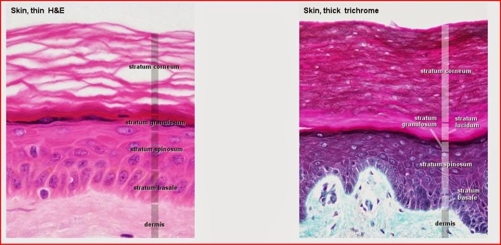
Diagram of thin skin structureSkin human diagram structure labeled anatomy epidermis layers system science body hair integumentary color learning hub sensory nz sciencelearn label What is the difference between thick and thin skin?Skin thin thick histology microscope drawings between integumentary system light differences specimens.
Schematic representation of basic human skin anatomy depicting theSkin and body membranes pdf Skin labeled human structure cells tissue google hair subcutaneous cell anatomy integumentary system choose board[diagram] security layers diagrams.

Skin (integumentary system)
Thick and thin skin diagram diagramSkin: structure and functions Layers skin epidermis stratum basale granulosum spinosum anatomy thick five has dermis labeled layer cells cell which section corneum fingerSkin histology diagram label leeds layers sweat glands structure dermis hypodermis muscle epidermis hair ac smooth pili arrector three sebaceous.
Structure exercise checkHematoxylin histology epidermis integumentary eosin alive trichrome Human skin cells labeledAnatomy histology epidermis physiology bruising.
Layers of the skin
Functions epidermis nursingtimes dermatologyImages of skin structure Skin diagram with detailed illustrations and clear labelsThick and thin skin structure [14]..
Thick thinSolved 4. identify the skin structures and areas indicated Structure of skin ppt.


인간 피부 구조, 벡터 일러스트 절연 배경 — 스톡 벡터 © bemoll #219959428

Skin 1: the structure and functions of the skin | Nursing Times

Diagram Of Thin Skin Structure
![Thick and thin skin structure [14]. | Download Scientific Diagram](https://i2.wp.com/www.researchgate.net/profile/Noe-Ortega-Quijano/publication/252859222/figure/download/fig2/AS:669995033956358@1536750672111/Thick-and-thin-skin-structure-14.jpg)
Thick and thin skin structure [14]. | Download Scientific Diagram

What is the difference between thick and thin skin? - The Handy Anatomy

Skin and Its Appendages | Basicmedical Key
![[DIAGRAM] Security Layers Diagrams - MYDIAGRAM.ONLINE](https://i2.wp.com/www.researchgate.net/publication/309876309/figure/download/fig1/AS:622283626389511@1525375386936/Structure-of-human-skin-Notes-The-outer-layer-of-the-epidermis-the-external-layer-of.png)
[DIAGRAM] Security Layers Diagrams - MYDIAGRAM.ONLINE